This is an online quiz called THS Anatomy Pelvis Posterior View. The two pelvic bones are connected anteriorly by the pubic symphysis while posteriorly they articulate with the pelvic spine to form the sacroiliac joints.
Muscles Of The Pelvis
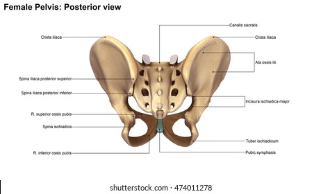
Major ligaments and notches of the female pelvis posterior view.

. The pelvis is composed of the two pelvic bones and the sacrum and coccyx. The Adult Heart- Anterior Surface View. The right and left hip bones also converge anteriorly to attach to each other.
The three bones and three joints composing the pelvic ring have no inherent stability without vital ligamentous structures. The pelvic spine is the posterior portion of the pelvis below the lumbar spine composed of the sacrum and coccyx. 184166848 stock photos online.
Manual Therapy for the Low Back and Pelvis A Clinical Orthopedic Approach 2015. From inception of the study to April 6 2018 MEDLINE database was used to search for 40 terms relevant to the posterior female pelvis and vulvar anatomy. Start studying Pelvic Bone Anatomy.
Adult Heart- Anterior View. The pelvic region of the trunk is the lower part of the trunk between the abdomen and the thighs. Identify the following parts of the pelvic girdle This quiz has tags.
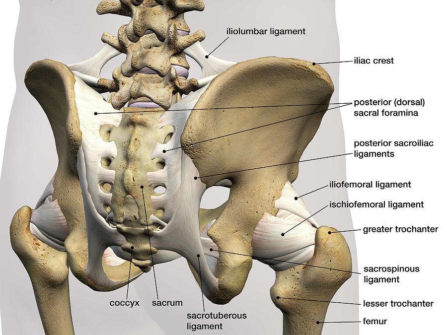
You may also find sacrospinous ligament lesser sciatic foramen sacrotuberous ligament ischial tuberosity deep posterior. E-FIG A2-3 Muscles of the pelvic diaphragm oblique view. This becomes important during parturition.
Evolutionary scientists believe this stems from mans hunter roots as a leaner pelvis made running easier. Click on the tags below to find other quizzes on the same subject. Cardiovascular System of the Lower Torso.
Pelvic Ligaments Biomechanics of the Pelvis. Lateral pelvic wall It is formed by. Learn vocabulary terms and more with flashcards games and other study tools.
From the quiz author. The pelvic bones are smaller and narrower. Pelvic ligaments posterior view.
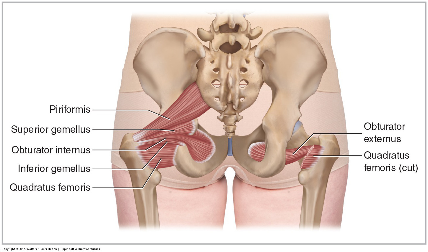
The quadratus femoris has been cut on the right side to visualize the obturator externus. The Cardiac Electrophysiologic Conduction System. New users enjoy 60 OFF.
The pelvis is a ring structure made up of three bones. Click on the tags below to find other quizzes on the same subject. The symphyseal ligaments which hold the pubis together resist external rotation and account for only 15 of the stability to the entire ring.
Most of which reflect the role of childbirth in the female. Skeleton Pelvis Posterior View. Anatomical landmarks within the vagina can be used to locate the position of such structures as the ureter and urethra and warn of their possible involvement in a vaginal laceration.
Download 1536 Posterior View Body Stock Illustrations Vectors Clipart for FREE or amazingly low rates. Part of the hip bone below the pelvic inlet the arrow 2. Bony pelvis is formed posteriorly by the sacrum and the coccyx and laterally and.
All muscles of the group are drawn on the left side the obturator externus is not seen. Identify the following parts of the pelvic girdle This quiz has tags. The parietal pelvic fascia is removed to visualize the embedded autonomic pelvic nerves.
The bones of the pelvis are the hip bones. Digestive System of the Lower Torso. During pregnancy temporary changes take place in the ligaments that permit both movement of the joints and enlargement of the pelvic cavity.
Start studying Female Pelvis - Posterior View. The male pelvis is smaller and narrower with a thinner pubic symphysis. Topographic anatomy of the posterior pelvic compartment.
De la prueba autor. The lumbar spine is composed of five vertebrae named L1 to L5 from superior to inferior. This is an online quiz called THS Anatomy Pelvis Posterior View.
The Adult Heart- Posterior Surface View. Each hip bone in turn is firmly joined to the axial skeleton via its attachment to the sacrum of the vertebral column. Posterior pelvic wall It is large and deeper.
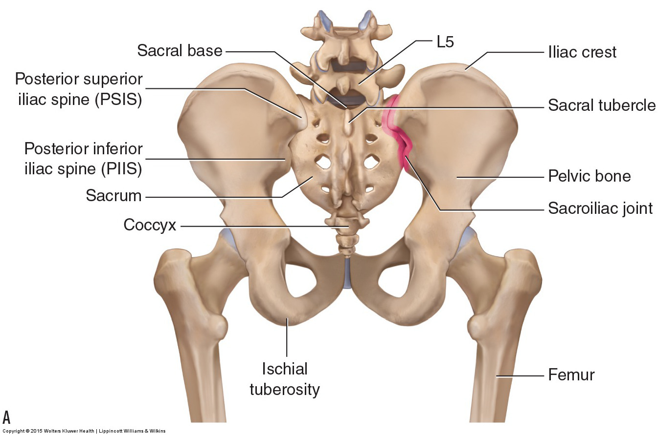
The vertebral column of the lower back includes the five lumbar vertebrae the sacrum and the coccyx. In this image you will find the posterior superior iliac spine iliac crest tubercle of the iliac crest anterior superior iliac spine greater sciatic foramen the acetabular margin in it. Posterior view of the deep lateral rotator group.
The pelvis is a ring structure made up of three bones. Learn vocabulary terms and more with flashcards games and other study tools. There is a printable worksheet available for download here so you can take the quiz with pen and paper.
Posterior view of the lumbar spine and pelvis. Abdominal Male Pelvic Anatomy- AP View. The posterior wall is next to the perineal body rectum and peritoneal cavity at the pouch of Douglas while the two lateral walls lie against the pelvic diaphragm and major vaginal vessels.
Manual Therapy for the Low Back and Pelvis A Clinical Orthopedic Approach 2015. The pelvis plays several important functions in the human body. The posterior ring structures are responsible for the majority of pelvic ring stability.
Bony pelvis or pelvic skeleton is formed by hip bones sacrum and coccyx. Download Human Skeleton System Pelvis Anatomy Posterior View Stock Illustration and explore similar illustrations at Adobe Stock. The female on the other hand has a much wider and more.
The sacrum and two innominate bones. Download scientific diagram Anatomy of the pelvis. There is a printable worksheet available for download here so you can take the quiz with pen and paper.
The Adult Heart- Long Axis Section. Bones of the Pelvis and Lower Back Posterior View Toggle Anatomy System. Bone And Ligaments Of Pelvis Posterior View.
Furthermore 11 investigators reviewed identified abstracts and selected those reporting on posterior female pelvic and vulvar anatomy for full-text review. 1 sacrum 2 coccyx 3 piriformis muscles right and left and 4 their covering of parietal pelvic fascia. The pelvic girdle hip girdle is formed by a single bone the hip bone or coxal bone coxal hip which serves as the attachment point for each lower limb.
A The posterior pelvic compartment is delimited from the urogenital compartment by the rectoprostatic septum Denonvilliers fascia. Medial view of a right-sided male hemipelvis.

Pelvis Anatomy Recon Orthobullets

Pelvis And Hip Anatomy Poster Pelvis Anatomy Anatomy Bones Hip Anatomy
Bones Of The Lumbar Spine And Pelvis

The Pelvic Girdle And Pelvis Anatomy And Physiology I
Skeleton Pelvis Posterior View 3d Illustration Stock Illustration 474011278
Male Hip Bones And Ligaments Labeled Rear View On Black Stock Photo Download Image Now Istock

Three Dimensional Posterior View Of The Pelvis Download Scientific Diagram
Rear View Of Male Pelvis Hip Leg Photograph By Hank Grebe
0 comments
Post a Comment